Scanning Electron Microscopy
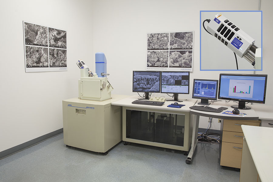
Our JEOL JSM-6610 Scanning Electron Microscope (SEM) has a large specimen chamber capable of handling a 200mm diameter specimen up to 65mm in height. Specimens are mounted on a 5-axis motorized sample stage with tilt and rotate capability. The SEM is equipped with an Oxford X-MaxN X-ray Energy Dispersive Spectrometer (EDS) for elemental analysis.
Secondary Electron Imaging and Backscatter Electron Imaging
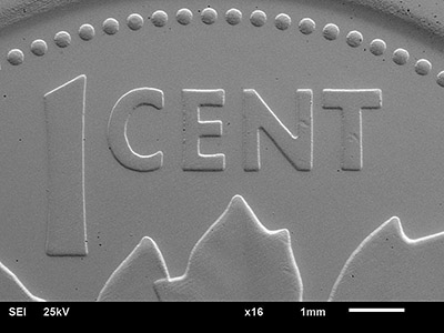
The SEM is capable of Secondary Electron Imaging (SEI) which is the "normal" image of surface texture. Backscatter Electron Imaging (BEI) shows a gray scale proportional to atomic weight of the elements in the field of view. BEI imaging is of particular interest for quickly finding flaws in material because it clearly shows variations in composition.
Imaging Service: we can prepare and photograph your sample, and prepare a PDF of image plates. Original JPEG photos can also be provided for use in your reports and projects. If required our clients can sit with the SEM operator to guide the image selection.
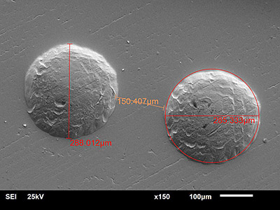
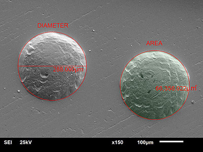
Measurements: the SEM can provide quantitative measurements of sample features. Annotated photographs of data are provided.
Sample Preparation: for optimal SEM imaging and higher magnification work the sample must be conductive. Non-conductive samples can be vacuum sputter coated with a 10Å thick layer of gold or carbon. Samples must be dry and not oily to avoid contamination of the SEM. We offer a sample cleaning / filtering service where necessary.